
The Receipt
Aquarius Mountains Garnet Series, Part 3

For those who are just joining, there is a garnet in my lab that I have been staring at for longer than I’d like to admit. It sits in a small tray. The face of it reflects light in a way that no garnet should — metallic green, iridescent, the color shifting slightly as the angle changes. Part 1 of this series explained where it came from and why the coating has been puzzling me since I first picked one up as a teenager in Mojave County, Arizona. Part 2 established, through X-ray fluorescence, that the coated garnets and the uncoated garnets at the same locality are chemically distinct populations — different trace-element signatures, different fluid histories, as if they had different pasts despite sharing the same outcrop.
This post is about what’s on the surface of the coated ones.
Not what the surface looks like, but what it is actually made of, and what that turns out to mean.
The Raman Question


When I examined the coating edge under the optical microscope, the boundary wasn’t a line. It branched. Fingers of material spread across the uncoated garnet face in a pattern that looked less like mineral film evaporating out of a solution and more like something advancing across a surface it hadn’t reached yet. Weathering rinds don’t have advancing fronts. They have boundaries that stop where the exposure stops. This one was still going somewhere, in a direction, with structure to the leading edge.
That was before any instrumentation. The shape of the question came before the answer.
Raman spectroscopy works like this: a laser fires at a sample, and almost all the light bounces back exactly as it arrived. But a tiny fraction comes back shifted — at a slightly different frequency. That shift happens because some of the laser’s energy was absorbed by the vibrating bonds inside the material and re-emitted. Different chemical bonds vibrate at different frequencies, so the pattern of shifts is a fingerprint. Not a rough one — specific enough to distinguish minerals that look chemically identical by almost every other method. On a coating this thin, the laser reads primarily what’s on the surface.
The uncoated garnet came back exactly as expected — a clean set of peaks belonging to almandine, the iron-rich garnet this locality produces. That’s the baseline. Everything else is what’s sitting on top of it.
Before we go further, a quick word about how to read a Raman spectrum, because the figure that follows is worth understanding. The horizontal axis is labeled in units called wavenumbers — a way of describing the frequency of the scattered light. You don’t need to know exactly what wavenumbers are. Think of the spectrum like a piano keyboard stretched out flat: different chemical bonds “play” different keys. Each mineral has its own chord — a specific set of positions where it shows up, and those positions don’t shift. The vertical axis just shows how loudly each key is being played.
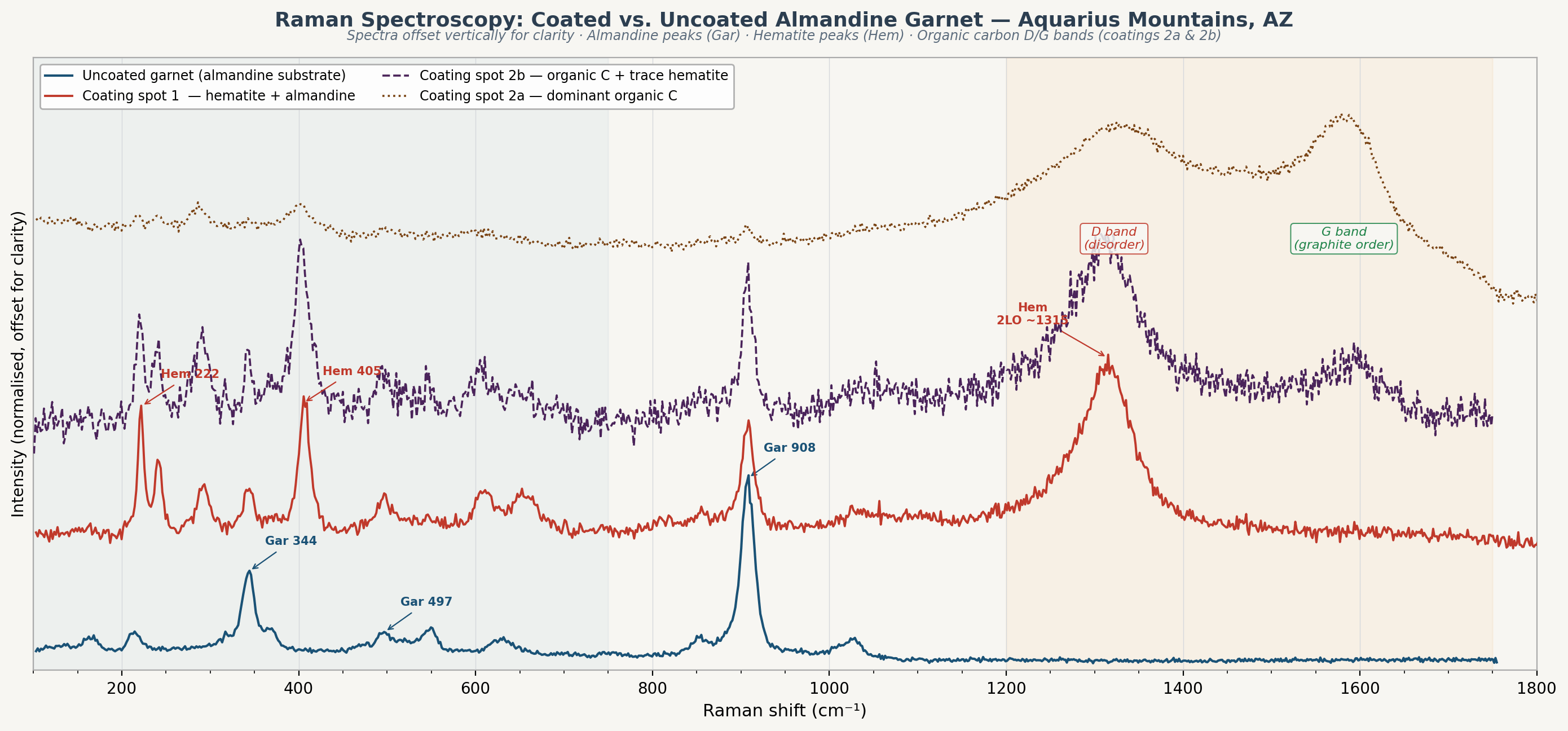
The coating spectra came back playing something I didn’t recognize.
Spot 1, measured at the coating margin, showed peaks characteristic of hematite — an iron oxide, the rust-red mineral that gives the Painted Desert its color and covers much of Mars. That is not a mineral you expect to find as a coating on a garnet. The garnet’s iron is in a form called ferrous iron — the stable, unoxidized form, the same kind you’d find in unrusted steel. The hematite in this coating carries ferric iron instead, the oxidized version. Rust, essentially. Something at the garnet surface converted one into the other, leaving hematite as evidence.
Here is where it gets slightly technical, but it matters, so let’s slow down. Hematite produces a faint extra peak at a position on the spectrum that happens to be very close to where organic carbon produces one of its characteristic signals. It is an overtone — the same phenomenon as the faint harmonic notes that ring alongside the main note when you pluck a guitar string. In spot 1, what might appear to be an organic signal is actually hematite playing one of its overtones. I am flagging this now because confusing the two would change the interpretation of the data, and I want the logic to be visible.
Spots 2a and 2b showed something else entirely. No hematite at all. Instead, two broad peaks in the high-frequency region of the spectrum — and these are not hematite. They are the signature of carbon.
Carbon atoms bonded to other carbon atoms in flat, orderly sheets — the arrangement you find in graphite — produce a signal called the G band. G for graphite. When that orderly structure breaks down — disordered, irregular, never fully crystalline to begin with — a second signal appears alongside it, called the D band, for disorder. The ratio of the two tells you how chaotic the carbon structure is. In pure graphite, D barely registers. Biological material is different: D tends to dominate, often louder than G, because living things don’t build their carbon in neat sheets.
In spots 2a and 2b, D dominates — broad, not sharp, clearly not graphite, not carbonate, not any form of carbon you would expect to find sitting on a garnet on a mountain in Arizona. It is disordered organic carbon. And it has not been cooked — the signal shows it is thermally immature, meaning whatever produced it was not buried deep or heated. It is relatively close, chemically, to what it was when it formed.
The coating contains hematite near the garnet surface, and organic carbon above it. Whether those are two distinct layers or one intermixed phase was not something surface Raman could resolve on its own. That required breaking it open.
The Plywood Discovery
LIBS — laser-induced breakdown spectroscopy — works by firing a high-energy laser pulse at the surface, ablating (vaporizing) a tiny amount of material. The resulting cloud of vaporized atomic plasma glows, and the instrument reads that glow. Different elements emit light at different wavelengths — the same reason fireworks are different colors — and that is how the instrument identifies what was there. Because each pulse goes slightly deeper than the last, LIBS is a depth-sampling technique: it reads the sample layer by layer from the surface down.
On a coating this thin, the first pulse or two consume it entirely and begin reading the garnet below. That was expected. What was not expected was what happened when I looked at the ablation crater — the tiny pit left behind — under the microscope afterward.
The coating hadn’t peeled away as a single unit. It had delaminated in layers — the way old plywood separates along its ply boundaries when water gets in. Two distinct layers, each lifting cleanly from the garnet below. The LIBS experiment had not characterized the coating chemistry the way I had hoped. What it had done, accidentally, was section it.
With the layers physically separated and the interfaces exposed, I pointed the Raman laser at each layer individually. The lower layer, sitting directly on the garnet surface, matched hematite. The upper layer matched disordered organic carbon.
The stratigraphy — the layered sequence of what came first, read from the bottom up the way geologists read rock strata — is: garnet → hematite (Fe₂O₃, iron bonded to oxygen) → organic carbon.
Three layers with different compositions, each confirmed by independent measurement, in a sequence with a direction. The hematite formed first, directly on the garnet surface. The organic carbon came after, on top of the hematite. Whatever process built this coating did it in order.
7,500×


The scanning electron microscope (SEM) tells the story in a different register. Unlike an optical microscope, which uses light, the SEM uses a focused beam of electrons to trace the surface of a sample. Because electrons have a much shorter wavelength than visible light, the SEM can resolve structure at scales thousands of times smaller than anything you could see with a conventional lens.
The coated face looks uniformly covered at low magnification — the metallic surface you see when you hold the specimen in your hand. Move up to medium magnification and the texture becomes irregular, lumpy, three-dimensional, nothing like the flat surface of the uncoated face beside it. At 7,500×, the structure finally resolves into something specific.
The coating is not a film. It is a packed array of sub-spherical (nearly round) structures, each roughly 0.5 to 2 micrometers across. For scale: a human hair is about 70 micrometers wide, so these structures are somewhere between one-thirtieth and one-tenth of a hair’s width. They sit against each other with no angular geometry, no flat faces, no straight boundaries. Rounded, packed, uniform in scale.
This is not what minerals look like when they grow. When inorganic compounds crystallize out of solution onto a surface, they produce forms that reflect their internal crystal structure — flat faces, angular boundaries, layered or branching patterns with geometric regularity. That is the signature of chemistry following crystallographic rules. Rounded, sub-spherical, non-angular structures packed at this scale are not a crystallization texture.
Coccoid bacteria — the sphere-shaped variety, the most basic bacterial form — are 0.5 to 2 micrometers across.
The resemblance is worth flagging. It is not a conclusion.
What the Beam Found
Energy-dispersive X-ray spectroscopy (EDS) works like this: when the electron beam strikes an atom in the sample, it can knock loose one of that atom’s inner electrons. When the atom refills that vacancy, it releases a small burst of X-ray energy. The energy of that burst is specific to the element — each element has its own signature X-ray, the way each element has its own spectral color in a flame test. By measuring the energies of all the X-rays coming off the sample, EDS identifies which elements are present and in what proportions. This specimen was run without any preparation coating — no carbon or gold applied to the surface before imaging. The numbers are from the sample itself.
The point measurement on the coating surface returned the following: carbon at 31.49% by mass (41.88% by number of atoms), nitrogen at 7.53% by mass (8.59% by atoms), oxygen at 38.55%, phosphorus at 2.08%, along with silicon, aluminum, calcium, potassium, sodium, and chlorine in smaller amounts. The two percentage figures for each element — mass% and atom% — describe the same thing in different ways: mass% is the proportion by weight, atom% is the proportion by count. Both are reported here because they tell slightly different parts of the story.
Three elements can be accounted for immediately: sodium, potassium, and chlorine. Those are sweat. I handled the specimen. Sodium chloride — table salt — and potassium are the chemistry of a fingerprint, not a garnet surface. Silicon, aluminum, and calcium are expected signal from the garnet itself bleeding through: at 15 kilovolts, the electron beam penetrates deeper than the coating is thick, sampling the almandine garnet below. Those elements belong to the garnet, not the coating.
That leaves carbon, nitrogen, oxygen, and phosphorus. Those are the coating.
The carbon is not a surprise. Raman already identified organic carbon in the upper layer. EDS confirms it and puts a number on it.
The nitrogen is what matters.
No mineral phase expected at this locality would produce a nitrogen signal. The contamination routes don’t work either — fingerprints don’t uniformly cover a surface, sample prep involved no nitrogen sources, and the atmosphere wasn’t a factor. Nitrogen at 8.59 atom%, alongside carbon and phosphorus, indicates that this coating contains organic matter of biological origin. In living things, nitrogen is the structural backbone of amino acids and proteins — the building blocks and machinery of cells. Phosphorus at 2.08% is the backbone of nucleic acids (DNA and RNA) and of phospholipid cell membranes, the fatty envelopes that contain every living cell. The co-occurrence of carbon, nitrogen, and phosphorus in these proportions is not ambiguous about what class of material is present.
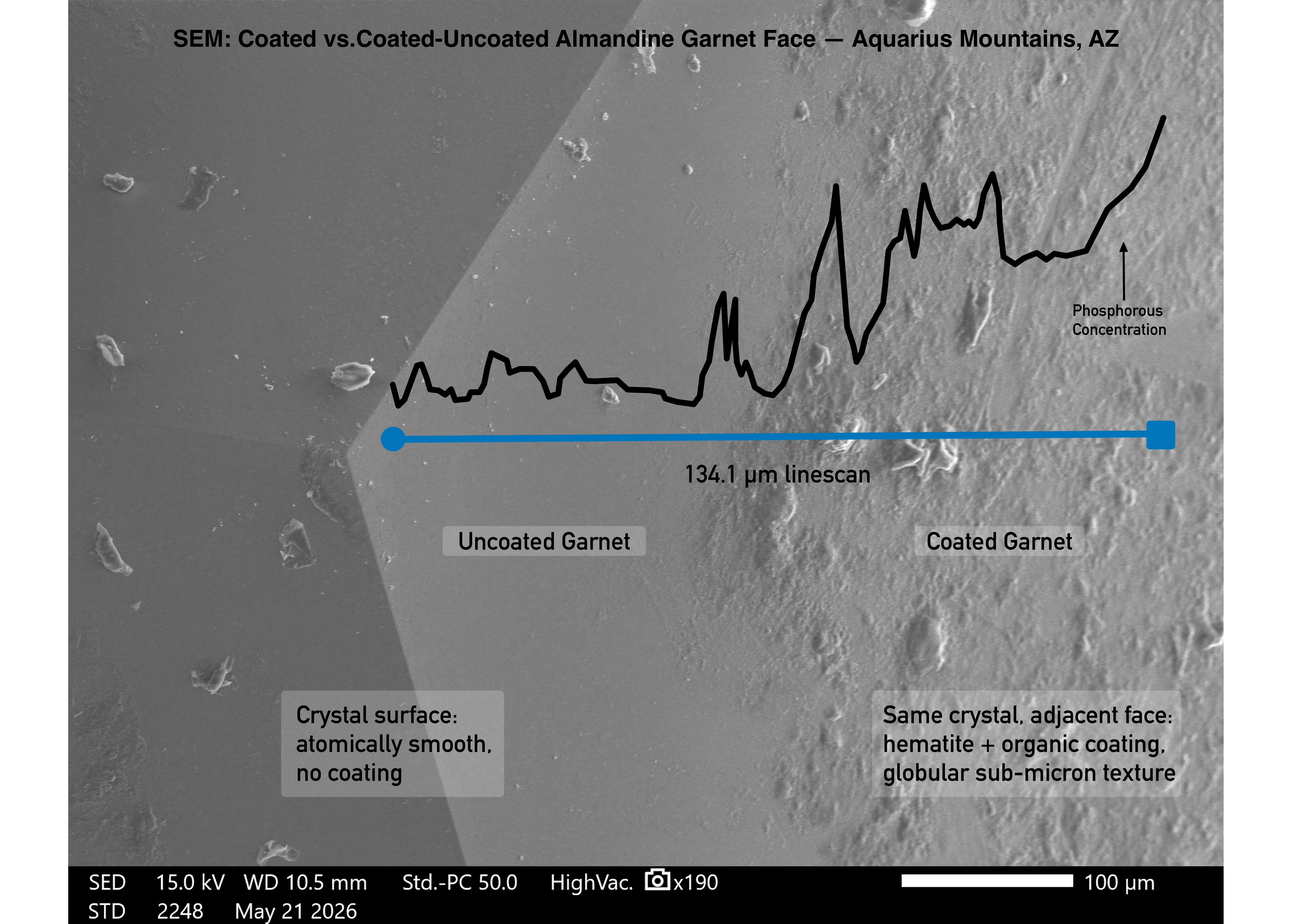
The EDS line scan at 134.1 μm and 190× plots phosphorus — carbon’s X-ray is too weak to map reliably at line scan acquisition speeds, so phosphorus serves as the spatial proxy. The signal runs low across the first half of the traverse (the uncoated garnet face), then rises sharply and remains elevated through the second half, with intense, irregular spiking. Carbon, calcium, and potassium follow the same trend. The rise marks the transition from uncoated to coated face. Within the coating zone, the spiking is irregular because the beam is crossing individual globules. Each spike is one structure. Phosphorus — which has no inorganic source at this locality — is not uniformly distributed across the surface. It is concentrated inside the globular domains, not in the spaces between them.
The carbon-to-nitrogen atomic ratio in this spectrum is approximately 4.9:1.
What the Sequence Means
The phosphorus resolves a question left open from Part 2. In the X-ray fluorescence (XRF) analysis in that post, phosphorus was elevated in the coated garnet population but ambiguous — the XRF beam is wide enough that it could have been reading the garnet itself, mineral inclusions inside it, or the coating. EDS can pinpoint the signal to specific locations on the surface, and it places the phosphorus in the coating. That changes what phosphorus means here. It’s in the coating, not the garnet below, and given everything else in that spectrum, the only reasonable description is biological.
The uncoated garnets from the same outcrop have none of this — no hematite layer, no organic carbon signal in Raman, nitrogen below detection. If this were simple weathering driven by exposure to air and water, both populations would show it. They live in the same mountain, experience the same conditions. Only the coated population shows the layered structure, and weathering doesn’t work selectively like that.
The most consistent interpretation of everything collected here is that iron-oxidizing bacteria — microorganisms that get their energy by converting ferrous iron (Fe²⁺) to ferric iron (Fe³⁺) — colonized the surface of these specific garnets, extracted iron from the crystal surface, and left hematite behind as the waste product of that process. The organic carbon layer above the hematite is what remained of the bacteria themselves: accumulated biomass that fossilized in place. The hematite isn’t incidental to the story. It is the receipt.
This is where the XRF chemistry from Part 2 becomes more than background. The garnet deposit occurs in organic-bearing host rock — the microbial community wasn’t introduced from somewhere else, it was already there, living in the surrounding material. What the XRF established is that the two garnet populations have different trace-element chemistries: the coated population carries elevated chromium, scandium, and zinc substituting into the crystal lattice, creating structural strain that makes its Fe²⁺ slightly less stable, slightly more available as an electron donor. The uncoated population, with a cleaner lattice, doesn’t offer the same opportunity. The bacteria didn’t colonize randomly. One population’s crystal chemistry made it worth the metabolic investment. The other didn’t. The coating is the record of that selection — not a weathering event, not something that arrived from outside, but the host rock’s own biology working on a specific subset of its own minerals.
There is a well-documented mechanism in Arizona desert mineralogy that may explain how the organic material got there in the first place. When a cactus dies, it doesn’t simply dry out and disappear. Its tissue is loaded with oxalic acid — a simple organic compound that cacti produce in large quantities during their lifetime — and as the plant decomposes, that oxalic acid and other organic compounds move downward through the soil and into the rock below. Mineralogist Anthony Kampf, Curator Emeritus at this museum, has spent years documenting what happens next: where those descending organic compounds meet iron-bearing and copper-bearing minerals in Arizona ore deposits, entirely new minerals form at the interface. Ferriphoxite, carboferriphoxite, and more than a dozen other new mineral species have been described from precisely this process at sites including the Rowley Mine in Maricopa County. It is a recognized pathway — surface biology writing a chemical record in the rock below.
The Aquarius Mountains garnets may be recording a version of the same story. A cactus — or many cacti, over time — died above this outcrop. Organic material moved downward. It contacted an iron-bearing mineral surface that was already in biological territory, already in contact with iron-oxidizing bacteria living in the organic host rock. What we measure in the coating is not directly what the cactus left behind. The C:N ratio of 4.9:1 tells us that. Cactus-derived oxalate contains no nitrogen — its C:N would be effectively infinite. Biological organic matter, the kind produced by living and dying microorganisms, runs between 4:1 and 6:1. Something processed the organic input. The coating is what that processing left behind: hematite as the metabolic product, biomass above it, the whole sequence preserved on the surface of a garnet that had the right chemistry to make it worth colonizing in the first place.
That interpretation is the most consistent reading of the data collected here. It isn’t a closed case. The sub-spherical morphology we see in the SEM is consistent with coccoid (sphere-shaped) bacteria and also consistent with other processes that produce rounded structures at that scale. The carbon-to-nitrogen ratio falls in the biological range, but it would also fall in that range for accumulated non-living organic material derived from biological sources. What would close the case — and what the instruments used here cannot provide — is molecular-level identification: lipid biomarkers (the distinctive fat molecules that form cell membranes), intact cell-wall chemistry, or preserved DNA.
What the instruments can say is that this coating has stratigraphy — a layered construction sequence running from garnet surface to hematite to organic matter — and that the organic matter contains nitrogen with no inorganic explanation, phosphorus that belongs in cell membranes and genetic material, and a carbon-to-nitrogen ratio of 4.9:1.
The garnet has been sitting in the collection drawer. The coating has been sitting on it. The question of what built it is still open.
Aaron Celestian is the Curator of Mineral Sciences at the Natural History Museum of Los Angeles County, a former scientist at NASA's Jet Propulsion Laboratory, and an adjunct professor at USC. The Aquarius Mountains garnets are part of the NHMLA research collection. He writes Pocketful of Χtals because mineralogy is stranger and more alive than most people have been told.
Fascinating. Do you think the bacterial activity and the resulting coatings developed over short (like thousands to tens of thousands of years) time frames, or over geologic spans, several million years? Or really, I'm asking how long ago it might have happened.
What a story! Thanks for bringing it to us Aaron!