
Something Chose This Garnet
Part 2: They look identical. The XRF says otherwise

I spent several weeks trying to figure out why a garnet would be green.
Not the crystal. The crystal, once you wipe the surface, is black. The green is on the outside — a film, nanometers thick in places, adhering to one population of garnets at this locality while leaving another population completely alone. Two garnets, side by side. Same habit, same hand-specimen color from a meter away — same approximate size if you were being careful about it. Put them under the microscope and one of them is doing something the other isn’t.


That asymmetry is what this post is about. Specifically: what an X-ray fluorescence spectrometer told me when I pointed it at both, and what it declined to answer.
What the Instrument Is Actually Doing
XRF works by firing X-rays at a sample and measuring the signal that comes back. Each element responds at a characteristic energy — iron at iron’s, calcium at calcium’s — and the instrument sums everything in the beam path into a single spectrum.
The beam on this instrument is 1.2 millimeters in diameter. It penetrates hundreds of microns into the sample. That matters here, because the coating on these garnets is extraordinarily thin — a film that produces optical interference colors, which means its thickness is on the order of visible light wavelengths: somewhere between 100 and 700 nanometers. A coating that thin is essentially invisible to XRF. The beam passes straight through it and reads whatever is below.
So when differences show up between the two populations, the coating isn’t what I’m reading. I’m reading the garnets.


I ran multiple measurements on each population. Seven spectra total — three on uncoated specimens, four on coated. The measurements are internally consistent within each population — which made the differences between populations harder to dismiss.
The Spectra Don’t Match
Here’s the spectral overlay. Blue is the uncoated population, averaged across three measurements. Orange is the coated population, averaged across four.
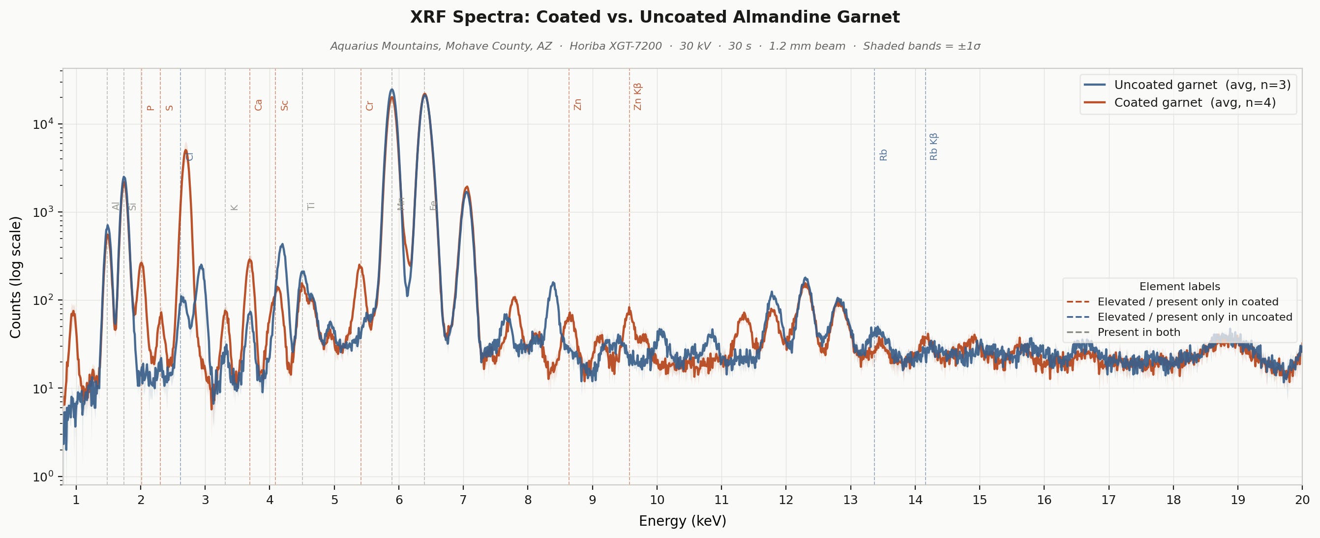
The dominant peaks — iron around 6.4 keV, manganese around 5.9 keV — are large in both spectra and broadly comparable. That's the garnet. Both populations are dominated by iron and manganese — the signature of Fe-Mn garnets in the pyralspite series (PYrope - ALmandine - SPessartine garnets). The Fe-to-Mn ratio differs between them, and that difference is one of the clearest signals in the dataset.
What’s different is everything else. Peaks around 2.0 and 2.3 keV are essentially absent in the uncoated spectrum; a feature at 3.7 keV that dwarfs the same region in the uncoated spectrum; a signal between 5.4 and 5.6 keV that the uncoated specimens don’t produce at all — four measurements, same result each time. Something is chemically distinct about these garnets.
The Complete Portrait of Population B
The quantified intensities put numbers to the spectral differences.
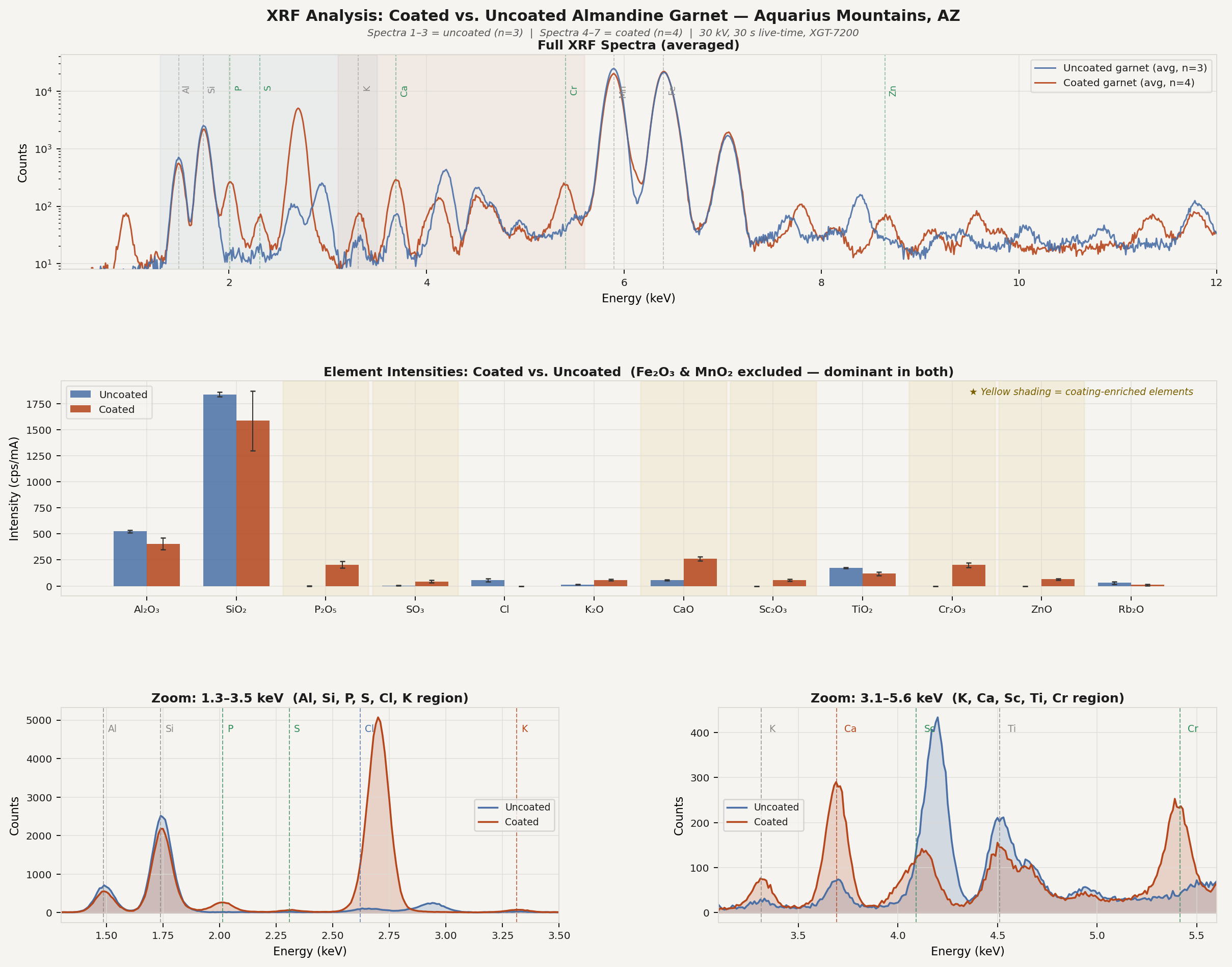
The uncoated garnets carry aluminum, silicon, chlorine, potassium, calcium, titanium, manganese, iron, and rubidium.
The coated garnets carry aluminum, silicon, phosphorus, sulfur, potassium, calcium, scandium, titanium, chromium, manganese, iron, zinc, and strontium. No chlorine. Rubidium either absent or dramatically depleted. K without Rb is a specific geochemical signature — more on that below.
Here’s what each element is telling me, because they are not all saying the same thing.
Phosphorus and calcium are elevated together — P at roughly 205 cps/mA in the coated population versus essentially zero in the uncoated population, and calcium roughly five times higher. Neither P nor Ca at these levels is surprising in the presence of apatite. Ca₅(PO₄)₃ is one of the most commonly documented accessory phases in vapor-phase and hydrothermal garnet assemblages. If Population B crystallized from a Ca- and P-bearing volatile phase that Population A never encountered, apatite micro-inclusions would explain both signals simultaneously.
Strontium tracks the calcium — 14 to 30 cps/mA across the four coated measurements, near zero in the uncoated population. Sr substitutes readily for Ca in apatite and Ca-bearing silicates. Its presence alongside the Ca anomaly is consistent: the same Ca-enriched volatile phase that produced elevated Ca would carry Sr in proportion. Sr has no structural site in almandine. Like K, it’s recording fluid history rather than garnet composition.
Sulfur is present at roughly 44 cps/mA in the coated population and only trace amounts in the uncoated. Sulfide phases are common accessories in vapor-deposited mineral assemblages in rhyolitic systems. But S can also concentrate in organic matter, and — I’ll say no more on this until the Raman post — there is reason to keep that possibility open.
Scandium at roughly 58 cps/mA in the coated population and zero in the uncoated is the most geochemically specific signal in the dataset. Sc has no structural site in almandine. It doesn’t concentrate in evolved felsic melts (think Mount St. Helens volcanic rocks) or sedimentary fluids in any meaningful amount. It partitions strongly into clinopyroxene in mafic rocks (think Hawaii-type volcanic rocks) and is mobilized by fluids that have equilibrated with those rocks. Its presence in Population B and its complete absence in Population A are strong evidence of different fluid-source chemistries.
Chromium at roughly 200 cps/mA in the coated, versus essentially zero in the uncoated, has the largest absolute difference among the diagnostic elements. Cr also has no structural site in almandine. Chromite (FeCr₂O₄), or chrome-spinel, as an accessory inclusion phase would point to a mafic component in the fluid or source-rock history. In a rhyolitic system where the volcanic suite runs from primitive basalt through to evolved rhyolites, a volatile phase that equilibrated with more mafic lithologies at any stage of that evolution could carry a Cr signature that a purely rhyolitic vapor would not.
Zinc at roughly 65 cps/mA in the coated versus zero in the uncoated. Zn is volatile-soluble and concentrates readily in magmatic vapor phases, particularly in evolved and rhyolitic systems. Zn-bearing accessory phases in vapor-deposited mineral assemblages are well-documented. Its presence in Population B alongside Sc and Cr points toward a vapor chemistry that Population A’s crystallization environment lacked.
P + Ca + Sr + Sc + Cr + Zn + S. Lined up like that, they tell a consistent story: Population B crystallized from a volatile phase that Population A never encountered. None of these elements has a structural site in almandine — they’re almost certainly sitting in accessory phases and inclusions, trapped during growth, recording the chemistry of that fluid.
The Fe and Mn signals deserve a direct comment, and they deserve it together because they are connected in ways XRF cannot fully resolve.
Mn averages roughly 27,150 cps/mA in the uncoated population and 21,850 in the coated — a 24% difference the coating cannot explain. That difference is in the garnets themselves.
The Fe picture is messier. The coated population averages higher but scatters widely across four measurements (20,800 to 26,150 cps/mA), while the uncoated population clusters tightly (21,932 to 22,472). That scatter is telling me something real about the coated garnets — but XRF has a specific limitation here that matters enormously for interpretation.
The leading hypothesis going into the next instrument is that the coated garnets are compositionally zoned — not homogeneous almandine, but almandine-dominant zones intercalated with zones carrying an almandine-spessartine component. Spessartine is Mn₃Al₂Si₃O₁₂; almandine is Fe²⁺₃Al₂Si₃O₁₂. If those zones are volumetrically minor relative to the almandine matrix, bulk Mn stays lower than in the uncoated population while the Fe signal scatters widely as the 1.2mm beam lands on different proportions of compositionally distinct zones across four measurements.
Why does the Fe/Mn ratio matter beyond accounting for the scatter? The coated population is more Fe-dominant — closer to pure almandine. The uncoated population is more Mn-dominant — closer to spessartine. Those are different surface chemistries. That difference may be exactly what the coating is reading.
That difference may be exactly what the coating is reading.
I will leave it there for now. The Raman post will say more.
K Without Rb Is a Very Specific Thing
Neither K nor Rb substitutes into almandine structure. Both signals are sitting in inclusions — K-feldspar, phlogopite, fluid inclusions — recording fluid history rather than garnet composition. Which makes the contrast between the two populations particularly telling.
The coated population has roughly four times more K than the uncoated, and essentially no Rb. The uncoated population has lower K and measurable Rb.
The K/Rb ratio is a standard geochemical discriminator between fluid sources. Primitive mafic rocks — basalt, mantle-derived melts — have high K/Rb ratios, typically around 1000, because Rb concentrates in felsic phases that are absent in primitive mafic systems. Evolved granitic and pegmatitic fluids have K/Rb ratios below 200, sometimes well below.
The coated population: mafic signature. The uncoated: more evolved, more felsic, K alongside Rb instead of K without it.
Two garnets, same locality, different fluid histories written into the trace elements. The coating finds one and leaves the other alone — and whatever it’s reading in the surface chemistry was decided when these crystals formed, not when the coating arrived.
Where the Elements Point
The Aquarius Mountains contain a mafic volcanic field. It is not a distant inference. The northern Aquarius Mountains volcanic field covers roughly 400 km² and includes basaltic flows and cinder cones dated to approximately 24–20 Ma. These basalts are primitive — high magnesium numbers consistent with direct mantle derivation — and the broader volcanic suite runs from them through latites and dacites to rhyolites, with K/Rb and Rb/Sr ratios that are distinctive and measurable at each compositional stage.
The garnets themselves are vapor-phase precipitates from a rhyolitic system. A volatile phase derived from a magma with any mafic input — mixing, contamination, or simply erupting through mafic crust — would carry Cr, Sc, and the high K/Rb character that Population B records. Population A, with its lower K/Rb and measurable Rb, appears to be a vapor pulse from a more purely evolved rhyolitic source. Two vapor pulses, two garnet populations, same cavity.
The spatial relationship between the volcanic field and the garnet locality needs field verification. The Aquarius Mountains are roughly 45 miles long, and establishing the fluid pathway between any mafic source rocks and the Lion Spring locality requires a map and boots on the ground, not just a range name. I am not claiming proximity. I am claiming that a mafic source consistent with the geochemical signal exists within the same mountain range, and that fieldwork now has a specific question to answer rather than a general region to survey.
That’s not a small distinction — going into the field with a specific geochemical target is different from going in to look around.
What the Microscope Added
Before the open questions, one more dataset.
The branching, dendritic front advancing into the bare garnet surface is not the morphology of mineral precipitation from solution, which tends toward uniform layers or crystallographic forms. It’s the pattern of systems growing under diffusion limitation — where the leading edge advances faster than the bulk material can supply. Two processes produce this morphology: diffusion-limited aggregation of large organic molecules in a viscous fluid, and biological colony expansion, specifically biofilm spreading across a substrate.


This is the fully coated face. The rainbow shimmer is thin-film optical interference. The coating is nanometers thick — in some patches perhaps 100 nm, in denser areas perhaps approaching 700 nm. This is not a crust, not a mineral precipitate in the conventional sense. It’s a film — and at the margin, it looks like something that spread.
What it’s made of is the subject of the next post. I will say only this: Raman spectroscopy found more than one phase in that coating, and at least one of them was not on my list of candidates going in. If you’re assuming iron oxyhydroxide, you’re not wrong — but you’re not finished either.
Where XRF Runs Out of Road
The instrument has a hard edge, and it’s worth knowing where it is.
XRF integrates everything in the beam path — garnet lattice, micro-inclusions, surface phases, fluid inclusions, all of it summed into a single spectrum. Phosphorus could be in apatite inclusions trapped during crystallization, or in the coating, or in fluid inclusions at grain boundaries. Chromium could be in chromite grains enclosed within the garnet, or concentrated at the surface. The Fe scatter across four measurements could be zoning — almandine-dominant zones intercalated with almandine-spessartine zones at a scale the 1.2 mm beam averages over rather than resolves. Each of these is a testable hypothesis. None of them can be distinguished from this data alone.
What it cannot tell me is how that iron is distributed spatially — which zones are Fe-dominant, which are Mn-dominant, and whether the coating preferentially covers one over the other.
That’s not a flaw in XRF. It’s a precise description of what it’s designed to do. Without it, there’s no chemical distinction to explain, no fingerprint to interpret, no reason to ask about iron oxidation states, no specific question to take into the field. The question doesn’t exist yet.
But XRF has now answered what it can answer. The question it leaves open — where exactly are these elements, in what phases, in what oxidation states, and how are they spatially distributed — requires tools that resolve chemistry at scales far below 1.2 mm.
Raman spectroscopy is next. It identifies mineral phases and organic compounds by their vibrational signatures and can target individual spots on a surface, separating coating from substrate and — as it turns out — revealing something about the internal structure of these garnets that the XRF beam was averaging over entirely. After Raman, SEM/EDS provides sub-micron spatial resolution, imaging the coating architecture in cross-section and identifying the chemistry of individual phases at the garnet surface and within the garnet interior. And field geology closes the loop on the fluid pathway: where, specifically, did these volatile phases come from, which rock did they equilibrate with, and why did one pulse grow a garnet that a later fluid would find and coat, while the other did not?
Each instrument resolves a different layer of the same question, and none of them alone gets there. The chain is the method.
The next post will cover the Raman and scanning electron microscopy data. I’ll warn you now: it does not simplify things.
Specimens from the Aquarius Mountains, Mohave County, Arizona. XRF collected on a Horiba XGT-7200, 30 kV, 30-second live time, 1.2 mm beam diameter.
Part 1 of this series: [link to Part 1]. Part 3 (Lasers): coming.
As the volatile fluids moved through the passageways in the rhyolite, could they chemically evolve and pick up a wider variety of elements? Since the coated garnets (Pop. 2) occupy the upper margins of the outcrop, it leads me to speculate that the initial volatile fluid pulse was simpler, depositing the Pop. 1 garnets. As the system expanded outward and upward, the fluid chemistry changed through prolonged interaction with the host rock.
Furthermore, the connection of Pop. 2 to a more mafic signature suggests that the initial phase of the eruption was purely felsic. As the eruption sequence progressed, it may have tapped deeper, more mafic layers of a zoned magma chamber (or experienced a late-stage mafic injection). Highly mobile fluids moving through this later cycle would then be naturally enriched in Scandium and Chromium, capturing that distinct signature in the Pop. 2 garnets.
Correction, May 31, 2026: This post and Part 1 refer to these garnets as andradite throughout. That identification was based on field observation — black, dodecahedral, volcanic context — not chemistry. The XRF data in this post, and Mindat's listing for this locality, are both consistent with almandine rather than andradite. The garnet identity question is now part of the open investigation. Relevant passages have been updated.